A new three-dimensional bio-printing technology opens up a new frontier to study micro-organs in the laboratory
05.06.2023
The impact that organoids have had on biomedical research and will have on translational research is enormous. At the expense of this impact, like all new generation systems, the use of organoids holds some limitations. Unlike what happens in a living system, organoids are developed in the laboratory by a self-assembly mechanism that usually forms into spheroidal structures, which cannot be easily controlled. This limit is because organoids are grown in the laboratory within inaccessible solid three-dimensional matrices (hydrogels), whose shape is difficult to modify.
Within the new study, entitled “Hydrogel-in-hydrogel live bioprinting for guidance and control of organoids and organotypic cultures” published in the journal Nature Communications, researchers have developed and tested a system capable of controlling the shape and therefore the cellular activity of different types of organoids. The work was conducted by a research group coordinated by Nicola Elvassore (Veneto Institute of Molecular Medicine - VIMM and University of Padua) and Anna Urciuolo (Pediatric Research Institute Città della Speranza - IRP and University of Padua), in collaboration with UCL and ShanghaiTech.
Here, researchers have developed a new three-dimensional biological printing technique capable of modifying the shape of the matrices to influence their growth, shape and therefore their activity. This technology, called hydrogel-in-hydrogel live bioprinting, can modify the environment surrounding organoids at any time during growth, thus opening a new perspective for the study of cellular activities in complex systems.
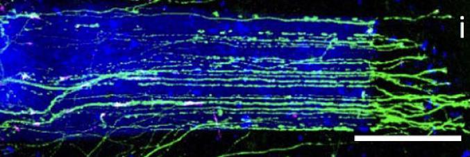
In particular, this study shows that it is possible to control the growth of spinal cord neuron extensions and better study its activity. Three-dimensional printing also allows for greater maturation of some cell types present in organoids, such as liver and intestinal. Furthermore, for the first time, researchers can control the growth of a three-dimensional model of the lung to mimic its morphogenesis during embryonic development. Finally, this technology will make it possible to study tumor organoids, opening up new perspectives in the field of stimuli that promote tumor cell migration, an activity involved in the possible formation of metastases.